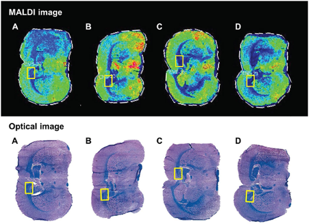
Figure 2. Histology directed MALDI-IMS analysis.
Brains from (A) control, (B) NNK-exposed, (C) ethanol-exposed, and (D) ethanol+NNK-exposed rats were sliced in the coronal plane to obtain a 3 mm slab that flanked the infundibulum of the hypothalamus. Adjacent cryostat sections were (Bottom) formalin-fixed and stained Luxol fast blue/Hematoxylin and Eosin (LHE), or (Top) mounted onto ITO-coated slides, sublimed with DHB and subjected to MALDI-IMS (see methods). In the LHE-stained sections, myelin is blue and gray matter structures are pink. The yellow rectangles represent co-registered regions of interest within the corpus callosum that were used for MALDI-IMS characterization of lipid ions.

