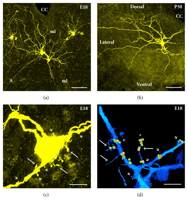
Figure 3.
Morphological properties of hypoglossal motoneurons in brainstem slices obtained from embryonic and adult C57/Bl6 wild-type mice (for methodology see Kanjhan and Bellingham, 2013 [26, 27]). (a) Image showing 4 hypoglossal motoneurons filled with Neurobiotin in a 300 μm slice preparation obtained from a mice at embryonic day 18 (E18). Note that the motoneuron on the dorsal right-hand side is dye-coupled to 4 adjacent motoneurons. Two motoneurons on the ventromedial portion of the hypoglossal nucleus have dendrites crossing the midline (ml) to the contralateral side. Axon (A) of one of the motoneurons is clearly visible projecting in the ventrolateral direction to join the hypoglossal nerve outlet. (b) A hypoglossal motoneuron from an adult mouse at postnatal day 30 (P30). Note a significantly larger dendritic tree in the adult mice. (c) A high-power confocal image showing filopodia (long arrows) and spine-like processes (short arrows) at the soma and primary dendrites of a motoneuron from a WT mouse at E18. (d) A rendered 3D reconstruction generated by Imaris software illustrating an overlapping localization of the presynaptic vesicular glutamate transporter-2 (VGLUT-2) terminals (small spheres) and the postsynaptic density protein-95 (PSD-95) (larger spheres) on filopodia (note as many as 4 excitatory synaptic contacts on a single filopodium marked as ∗) and spine-like processes on the primary dendrites of a motoneuron from an E18 WT mouse. CC: central canal. Scale bars = 100 μm in (a and b); 10 μm in (c and d).

