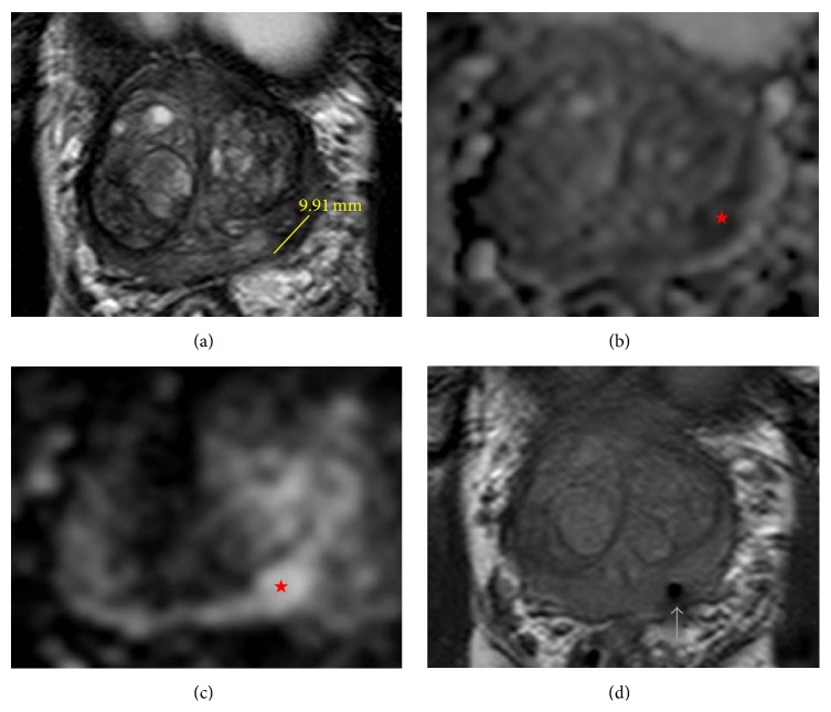
Figure 1.
(a) Magnetic resonance T2 weighted axial image of the prostate tumor (left base of the peripheral zone) with line indicating a tumor diameter of 9.19 mm. (b) Magnetic resonance diffusion weighted with apparent diffusion (ADC) mapping; lesion indicated by red star. (c) Magnetic resonance diffusion weighted with B2000 axial image of the prostate tumor (left base of the peripheral zone) indicated by the red star. (d) Magnetic resonance T2 weighted axial image confirming accurate needle placement in lesion prior to taking biopsy (arrow pointing to needle tract).

