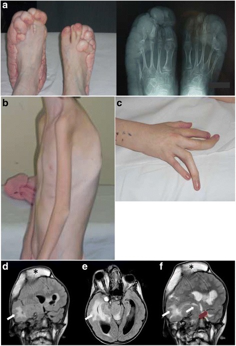
Fig. 1.

Clinical and radiologic presentation of the index PS patient. a Overgrowth of the feet (left panel) with radiologic confirmation of bone overgrowth, especially of the left foot (righ panel). b Severe cyphoscoliosis of the index patient. c Length discrepancy and macrodactyly in the III and IV fingers of right hand from the index patient. d e and f Coronal flair (d) and axial flair (e) weighted images demonstrated hyperostosis of the right fronto-parietal cranial vault (black asterisk) with proptosis of the right eye and extensive malformation of cortical development involving predominantly the right cerebral hemisphere but also part of the left cerebral hemisphere. There is an extensive white matter signal abnormality of both hemispheres (white arrows); there is also an associated malformation of the brainstem. Coronal T2 weighted images (f) show hyperostosis of the right fronto-parietal cranial vault (black asterisk), extensive malformation of the right hemisphere with white matter changes in the periventricular region (white arrows); malformation of cortex in the left perisylvian region (red arrow)
