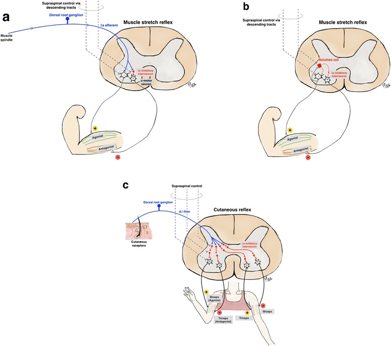
Fig. 1.

Major spinal reflexes include muscle stretch and cutaneous reflexes. a. The muscle stretch reflex. The afferent signal of the muscle stretch reflex from the muscle spindle is conveyed via Ia afferent fibers which are axons of dorsal root ganglia. The proximal axons then synapse directly with the alpha-motor neuron (a monosynaptic reflex), which in turn conveys an efferent signal to the corresponding extrafusal fibers of the agonist muscle leading to muscle contraction. Ia afferent fibers also synapse with Ia inhibitory interneurons, which in turn convey inhibitory signals to antagonist muscles, (reciprocal inhibition). b. The feedback control of the muscle stretch reflex. The Renshaw cell is the specialized inhibitory interneuron that functions in feedback control of the alpha-motor neurons in A. It receives input from collateral axons of the alpha-motor neuron controlling agonist muscles, and sends inhibitory signals back to the same alpha-motor neuron and also inhibitory signals to the alpha-motor neuron controlling antagonist muscles. The alpha-motor neurons, inhibitory interneurons and Renshaw cells also receive supraspinal control via descending tracts such as corticospinal or rubrospinal pathways. c. The cutaneous or flexion-withdrawal reflex. The cutaneous or flexion-withdrawal reflex is a polysynaptic reflex. Cutaneous nociceptive receptors send afferent signals via Aδ fibers which synapse with multiple interneurons before finally synapsing with the alpha-motor neuron. The interneurons connect the afferent and efferent signals, resulting in an excitatory signal to the ipsilateral flexor and contralateral extensor muscles, and an inhibitory signal to the ipsilateral extensor and contralateral flexor muscles. By this mechanism, flexion of the ipsilateral agonist muscle withdraws the limb from the nociceptive stimuli. An opposite chain is reversed in the opposite limb to prepare for support
