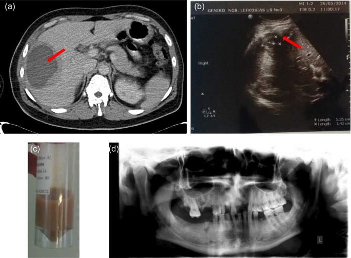
Figure 1:
(a) Abdominal CT scan with intravenous contrast showing a sizable (cyst containing) hypodense lesion with thickened wall (15 mm) in the right liver lobe (liver section 5 and 6), peripherally to the liver's capsule (red arrow). Lesions maximum dimensions were 10 × 10 × 8 cm. It displayed also minimum fluid collection around the liver. The imaging findings were primarily suggestive of an abscess. Another possibility would be a tumor with central necrosis. Additionally, there were small para-aortic lymph nodes with diameter <1 cm as well as mild splenomegaly (spleen diameter 13.5 cm). (b) Follow-up ultrasonography imaging a week after having the drainage catheter in situ: residual abscess (red arrow) cavity with dimensions of 5.35 × 1.42 cm with minimal effusion and air. (c) Light-brown abscess fluid sample, after CT-guided drainage. (d) Patient's panoramic radiograph with the absence of abscesses.

