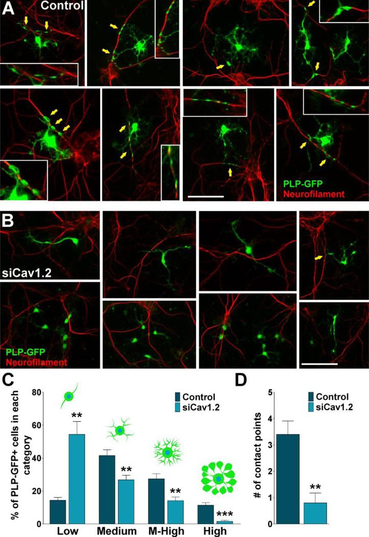
FIGURE 8. Co-culture of cortical neurons and VOCC deficient OPCs.
(A–B) PLP-GFP labeled OPCs were transfected with siRNA duplexes specific for Cav1.2 (siCav1.2). 24h after siRNA transfection, control and transfected OPCs were co-cultured with cortical neurons for 7 days. Arrows indicate oligodendrocyte-neurite contact points (see insets for high magnification pictures). Scale bar = (A) 60µm; (B, upper panel) 60µm, (B, lower panel) 80µm. (C) Morphological complexity of control and siCav1.2 transfected PLP-GFP+ cells in co-culture was scored in 4 categories. (D) The number of contact points with neurites was analyzed in control and Cav1.2 transfected cells. Results are the means ± SEM for three independent experiments. >200 cells were analyzed per experimental condition.**p<0,01 and ***p<0,001 vs. respective controls.

