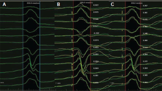
Figure 2: Pace Mapping of a Ventricular Premature Beat Utilising the PaSo® Module of CARTO® System.

A. The mapped ventricular premature beats (VPBs) have a left bundle branch block (LBBB) morphology and inferior axis, transition zone in lead V4, indicating an origin from the right ventricular outflow tract. B. During pacing over the mapping catheter at a free wall site with a cycle length of 600 milliseconds, a paced QRS morphology was produced. The latter was superimposed onto the native QRS morphology and the the PaSo® Module calculated the correlation between paced and native QRS morphology in each lead. Finally, an overall correlation percentage was demonstrated, in this case 87 %, driven mainly by the positive lead aVL on the pacing site, in contrast to the negative aVL of the native QRS. C. Pacing at a more septal and superior site produced a correlation of 0.97 %. Ablation at this site successfully terminated the VPB.
