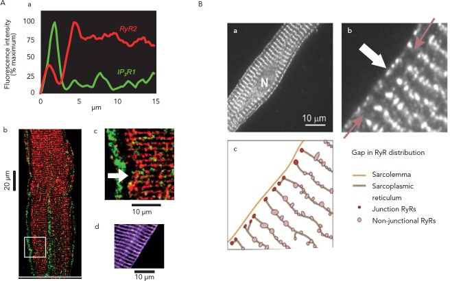
Figure 2: Architecture of Ca2+ Release Channels in Purkinje (A) and Atrial (B) Cells.
The Ca2+ release channels (RyR) are organised into clusters (red dots in A.b and A.c); the clusters distribution follows a transverse striated pattern that matches the striation of the contractile filaments (not shown here). In Purkinje cells, junctional RyRs co-localise with IP3 receptor channels under the membrane. Note that both cell types show a gap in the RyR2 distribution. The gap is absent when isoform non-specific RyR antibody is used (A.d), indicating the presence of a different RyR isoform in the ‘gap’ of Purkinje cells. The same RyR2 organisation is found in atrial cells (B.a, B.b) wherein the same gap is interpreted here as a space filled by sarcoplamic reticulum with no channel and separating ‘junctional’ and ‘Non-junctional’ RyRs (B.c). In both cell types, this microanatomy shown schematically in B.c sets the stage for successful Ca2+ wave propagation. Adapted from Boyden et al.,7 Thul et al.8 and Stuyvers et al.19

