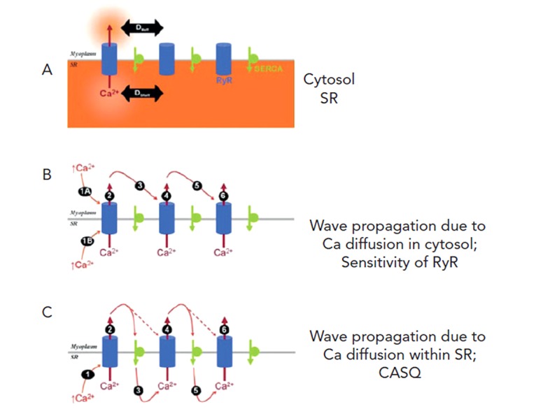
Figure 3: Simple Schematic Showing the Important Components of Ca2+ Wave Propagation.

Panel A: Elements included are RyR Ca2+ release channel (blue), SERCA pump (green) both in the sarcoplasmic reticulum (SR) membrane that separates myoplasm and SR lumen (orange). Panel B: Ca2+ in myoplasm (step 1A) acts on first RyR cluster to open channel to release Ca2+ stored in SR to the myoplasm (step 2). This Ca2+ then diffuses away (step 3) and interacts with anatomically close RyR cluster (see Figures 2B and C) to cause further CICR (step 4). This process is regenerative and relies on sensitivity of RyR to Ca2+. Panel C: Ca2+ wave propagation as in Panel B but now wave propagation is also occurring in SR lumen. This process is thought to depend on calsequestrin (CASQ). Adapted from Swietach et al.12
