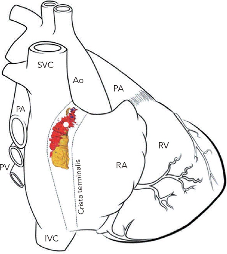
Figure 2: 3D Computer Reconstruction of the Human Heart Sinoatrial Node from Histological and Immunohistochemical Data Demonstrating the Extent of the Sinoatrial Node and Peripheral Pacemaking Tissue.
Red = sinoatrial node (SAN); yellow = peripheral pacemaking tissue; the leading pacemaker is shown as a white dot in the superior aspect of the SAN. Ao = aorta; SVC = superior vena cava; PA = pulmonary artery; PV = pulmonary vein; CS = coronary sinus; IVC = inferior vena cava; RA = right atrium; RV = right ventricle. Adapted from Chandler et al.84 Reproduced with permission from Wiley-Liss, Inc.

