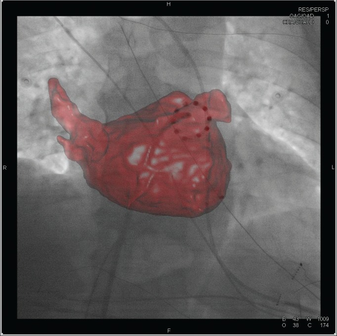
Figure 1: 3D Reconstruction of Left Ventricular and Pulmonary Vein Anatomy from Rotational Angiography.

The 3D reconstruction is superimposed on a live radiographic image. The reconstruction is used to guide manipulation of a circular multi-electrode catheter which is positioned at the ostium of the left inferior pulmonary vein.
