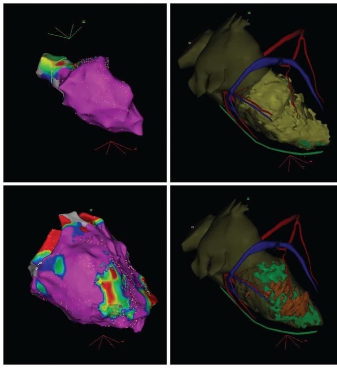
Figure 3: Pre-procedural Imaging in a Patient with Previous Myocarditis Presenting with Ventricular Tachycarida.

A) Endocardial and epicardial substrate maps. Purple demonstrates normal tissue (>1.5 mV bipolar voltage). Endocardial substrate map demonstrates absence of scar. Epicardial map demonstrates a small inferior lateral scar which could potentially have been missed in the absence of pre-procedural imaging. B) Multidetector CT (MDCT) segmentation of left ventricular endocardium and epicardium. Wall thinning is illustrated in green (<4 mm) and orange (<2 mm). Anatomical structures such as the coronary sinus (blue), coronary artery (red) and phrenic nerve (green) are also indicated. C) MDCT and late gadolinium enhancement MRI fusion image with MRI scar demonstrated in yellow. Coronary sinus is depicted in blue, phrenic nerve in green and coronary artery in red.
