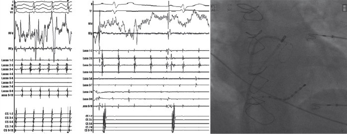
Figure 1: Pulmonary Vein Trigger Driving Atrial Fibrillation.
A: Intracardiac recordings from a circular mapping catheter (lasso 1–2 – lasso 9–10) positioned in the left inferior pulmonary vein (LIPV). The recordings demonstrate rapid activity which is conducted to the atrium and drives a rapid atrial arrhythmia. Atrial signals are recorded in a decapolar catheter positioned in the coronary sinus (CS1–2 – CS9–10). RFp and RFd demonstrate signals recorded from an ablation catheter which is being used to electrically isolate the PV. The surface electrogram is demonstrated on the top. B: Following isolation of the LIPV, the vein continues to fire rapidly, however the impulses are not conducted to the left atrium. The atrium is in normal sinus rhythm. C: A radiographic image (anterior–posterior projection) demonstrating position of the circular mapping catheter in the LIPV, the ablation catheter at the ostium of the PV and the CS catheter in the coronary sinus.

