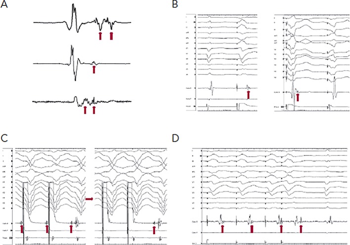
Figure 1: Important Mapping Concepts.

A) Sinus rhythm electrograms (EGMs) demonstrating late potentials and multicomponent EGMs (arrows). B) Right ventricular pacing utilised to expose late potentials (arrows) hidden within the QRS during sinus rhythm (left panel) and biventricular pacing (right panel). C) Entrainment mapping with capture of the far field EGM (left panel). A reduction in pacing output (right panel) results in successful capture of the local EGM (arrow) and evidence of concealed entrainment. D) EGMs recorded from abnormal substrate are poorly coupled to the rest of the myocardium. RV pacing results in separation of the late potential (hidden within the sinus QRS beat), and extrastimulus results in further delay.
