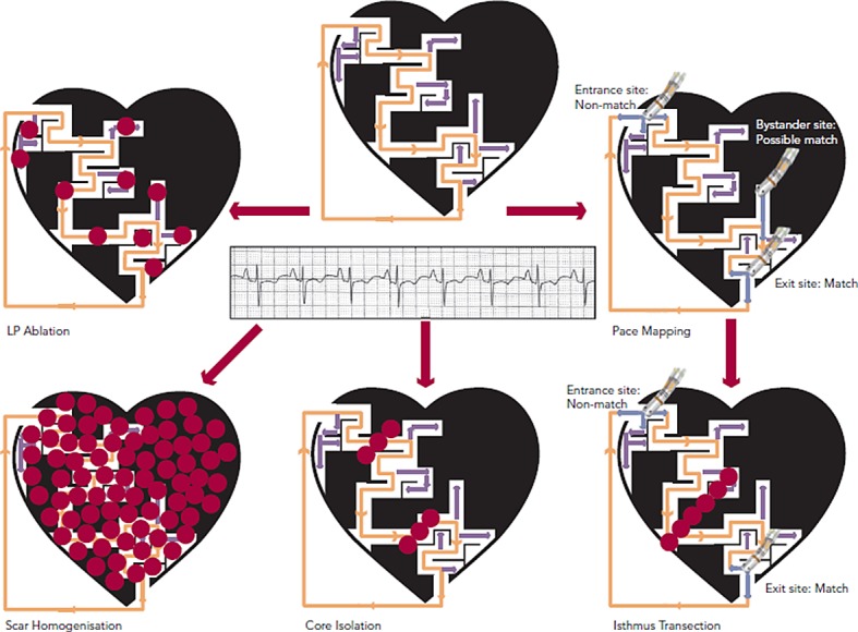
Figure 4: Mapping and Ablation During Normal Sinus Rhythm.

LP= late potentials. Dense scar is depicted in black with a conducting channel of viable myocardium. Inactive VT circuit is depicted with orange arrows and bystander areas with light purple arrows. Different approaches to mapping and ablation include LP ablation, pace mapping with isthmus identification and transection, scar homogenisation and core isolation. Ablation (red dots) is targeted at interrupting the VT circuit.
