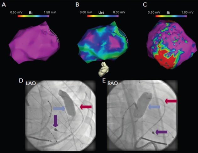
Figure 5: Electroanatomic Mapping in a Patient with Ventricular Tachycardia and Underlying Dilated Cardiomyopathy.

Endocardial mapping reveals normal bipolar (panel A) but abnormal unipolar voltage (panel B), suggestive of mid-myocardial or epicardial scar. Epicardial bipolar mapping (panel C) reveals extensive scar substrate. Due to phrenic nerve capture in this area, inflation of a contrast-filled balloon (blue arrow) in the epicardium was utilised to separate the phrenic nerve from the epicardial surface, with the ablation catheter (red arrow) in contact with the epicardium below the balloon (panels D and F). A left ventricular assist device was used to facilitate mapping and ablation (purple arrow).
