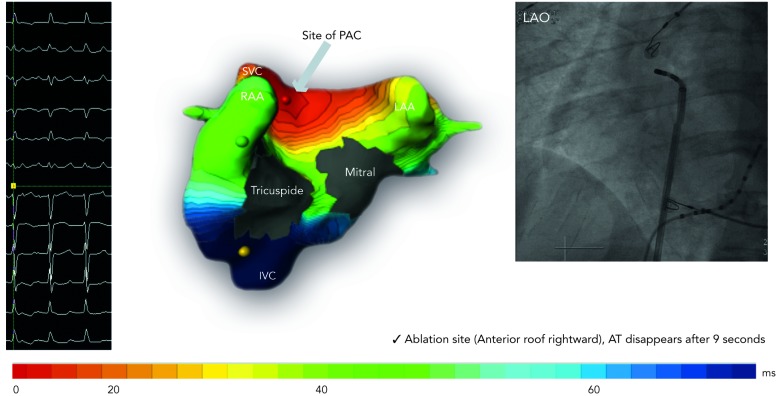
Figure 5: Focal Source Premature Ectopic Complex Arising from the Anterior Wall of the Right Superior Pulmonary Venous Ostium was Mapped Accurately with the ECM System (Isochronal Map Shown) and Successfully Ablated at the Corresponding Site Seen on the Fluoroscopic Image. Premature Ectopic Complex Terminated After 9 Seconds of Radiofrequency Application.
AT = atrial tachycardia; IVC = inferior vena cava; LAA = left atrial appendage; LAO = left anterior oblique; ms = milliseconds; PAC = premature atrial complex; RAA = right atrial appendage; SVC = superior vena cava.

