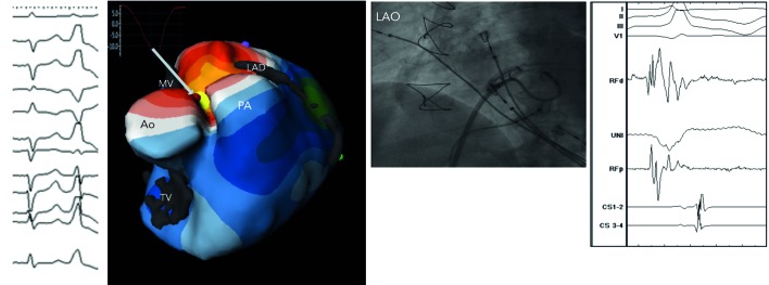
Figure 6: Biventricular Isopotential Map (Yellow Colour Denotes the Earliest Activation) During Premature Ventricular Complex (12-Lead ECG). Inserted is an Epicardial Virtual Electrogram (QS Morphology) from the Earliest Site. The Fluoroscopic Image at the Site of Successful Ablation and Local Intracardiac Electrogram are Shown As Well.
Ao = aortic root; LAD = left anterior descending artery; LAO = left anterior oblique; MV = mitral valve; PA = pulmonary artery; TV = tricuspid valve.

