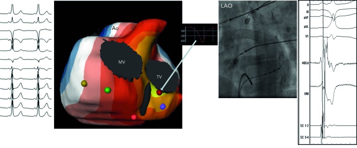
Figure 7: Biventricular Isopotential Map (Yellow Colour Denotes the Earliest Activation) During Pre-excitation from a Right Posteroseptal Accessory Pathway (12-lead ECG), which was Successfully Ablated (Fluoroscopic Image) at the Corresponding Site. Also Inserted is a Virtual Electrogram (QS Morphology) from the Site of Earliest Ventricular Activation Over the Manifest Pathway.
AO = aortic root; LAO = left anterior oblique; MV = mitral valve; TV = tricuspid valve.

