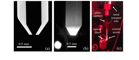
FIG. 2.
(a) The cross section of a ceramic injector tip obtained through x-ray tomography, which shows the 30° convergent cone and exit orifice of 100-μm diameter. (b) Image of the ceramic injector tip. (c) Image showing the nozzle mounted to the transport tube in vacuum. The end of a 400-μm-diameter fiber optic used for illuminating particles is shown to the left of the injector tip.

