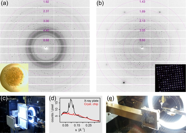
FIG. 2.
(a) X-ray diffraction pattern of a 40–50 μm T. daniellii thaumatin crystal located on a commercially available 96-well X-ray plate. The inset shows a photograph of the 1 μl crystallization droplet on the plate. (b) X-ray diffraction pattern of a 40–50 μm thaumatin crystal mounted on a crystallography chip with 50 μm sized features. The inset is a photograph of one compartment with 144 features at prescribed positions. The photograph in (c) shows the X-ray plate mounted at I24, Diamond Light Source. (d) Background scatter, radially averaged, as function of s = sinθ/λ. The photograph in (e) shows the crystallography chip mounted in its holder at beamline I24.

