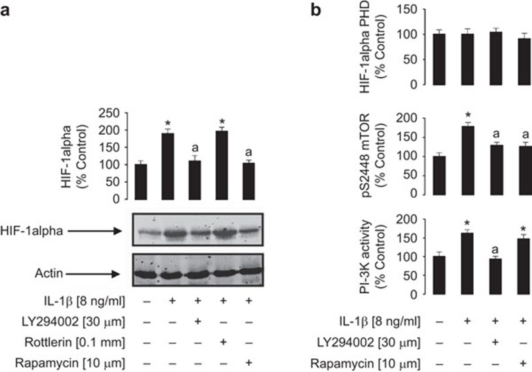
Figure 5.
The PI-3K/mTOR pathway is crucially involved in IL-1β-induced HIF-1α accumulation in MCF-7 cells. Cells were pre-treated with inhibitors for 1 h (as indicated) and subsequently exposed to 8 ng/ml IL-1β for 4 h. The procedure was followed by Western blot analysis of HIF-1α accumulation (a) and detection of PI-3K and HIF-1α PHD activities as well as phospho-S2448 mTOR deposition (b). HIF-1α Western blot data show one representative experiment of three similar results and were quantitatively analysed. Quantitative data are shown as mean values±s.d. of at least three individual experiments; results represent percentage values compared to the control. *P<0.01 vs. control; aP<0.01 vs. IL-1β treatment. HIF, hypoxia-inducible factor; mTOR, mammalian target of rapamycin; PHD, prolyl hydroxylase; PI-3K, phosphatidylinositol-3 kinase.

