Figure 1.
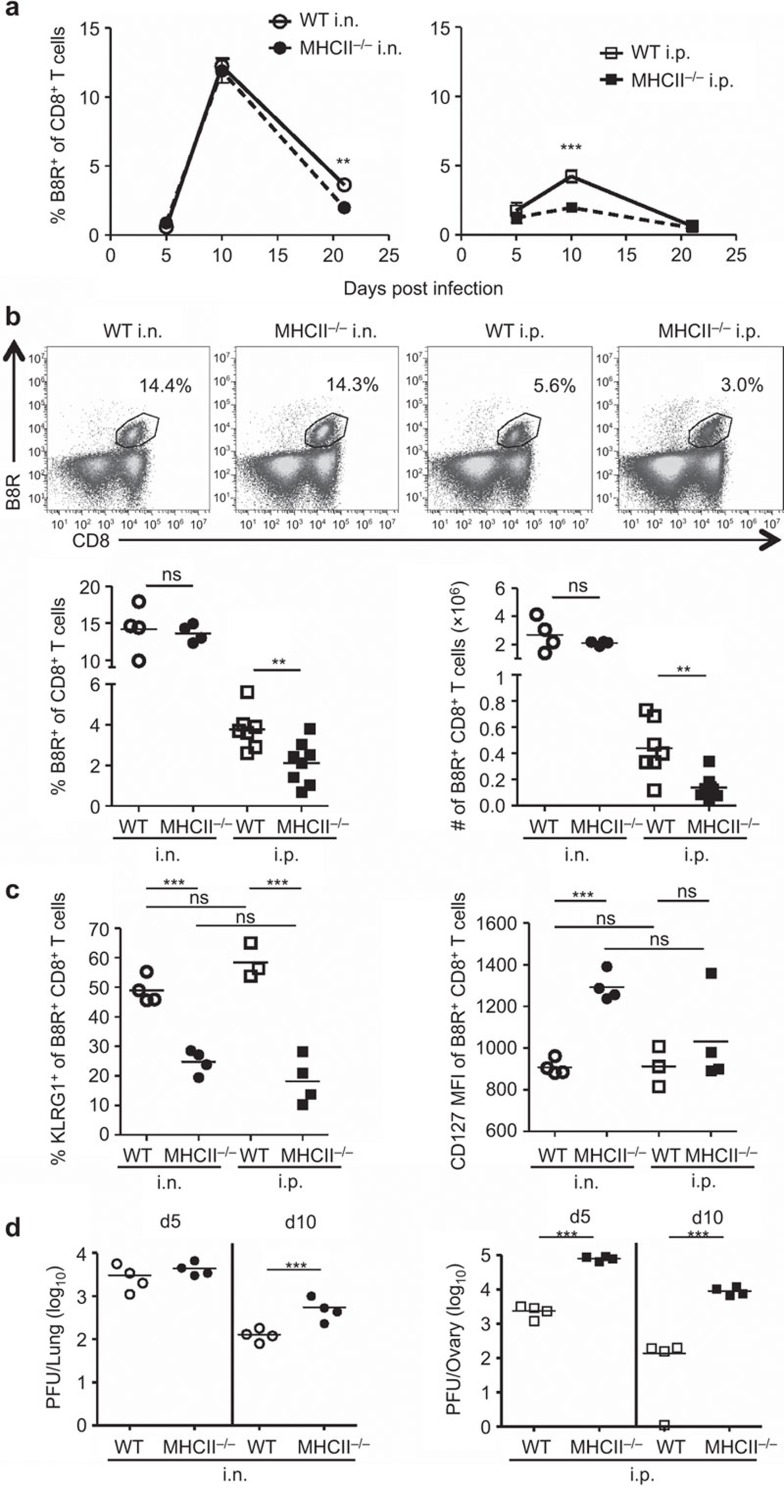
Primary CD8+ T-cell responses in intranasally or intraperitoneally infected mice in the absence of CD4 help. C57BL/6 (WT) and MHC class II knockout (MHC II−/−) mice were infected with 103 PFU of VACV-WR i.n. or i.p.. B8R-specific CD8+ T cells were identified by staining with B8R tetramer and anti-CD8α Ab at the indicated times. (a) Kinetics of B8R-specific CD8+ T-cell response. Frequencies of B8R-specific cells within the CD8+ T-cell population in the spleens of WT and MHC II−/− mice infected i.n. or i.p. are shown. (b) B8R-specific CD8+ T-cell populations in the spleens at day 10 post infection (pi). Representative FACS plots (top panel); frequency and total numbers (bottom panel). (c) Expression of KLRG1 and CD127 on B8R-specific CD8+ T cells at d9 pi. (d) Viral titers in lungs and ovaries, determined by plaque-forming assays. Each point on the graph represents a single mouse, and horizontal bars indicate the means. Data are representative of two to three independent experiments with three to eight mice per group. **P<0.01; ***P<0.001. Ab, antibody; i.n., intranasally; i.p., intraperitoneally; ns, not significant; pi, post infection; VACV-WR, vaccinia virus Western Reserve; WT, wild-type.

