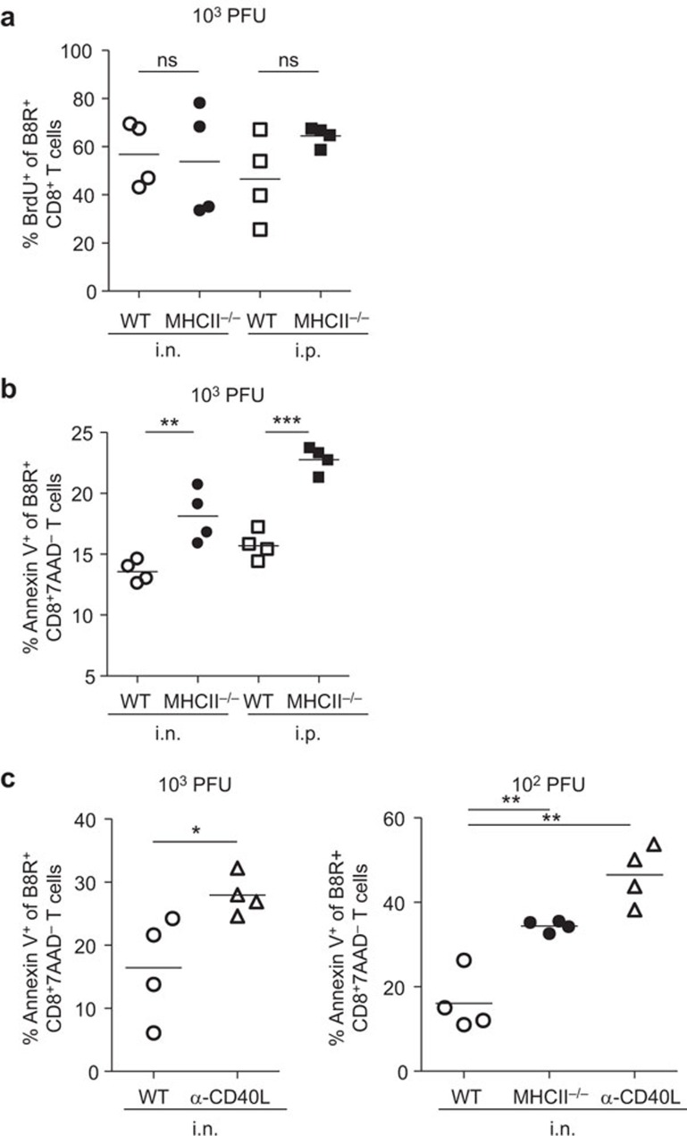
Figure 7.
Proliferation and apoptosis of VACV-specific CD8+ T cells. In vivo proliferation was measured by detecting incorporation of BrdU. Apoptosis was measured by Annexin V staining in conjunction with 7-AAD. B8R-specific CD8+ T cells were prepared from the spleens of infected mice. (a) Frequency of BrdU+ cells within B8R-specific CD8+ T-cell population in WT or MHC II−/− mice at d9 pi. (b) Frequency of Annexin V+ cells within B8R-specific CD8+7AAD− T-cell population in WT or MHC II−/− mice at d10 pi. (c) Frequency of Annexin V+ cells within B8R-specific CD8+7AAD− T-cell population in WT, MHC II−/− or α-CD40L WT mice at d9 pi. Data are representative of two independent experiments with four mice per group. *P<0.05; **P<0.01; ***P<0.001. 7-ADD, 7-aminoactinomycin D; BrdU, 5-bromo-2′-deoxyuridine; ns, not significant; pi, post infection; VACV, vaccinia virus; WT, wild-type.

