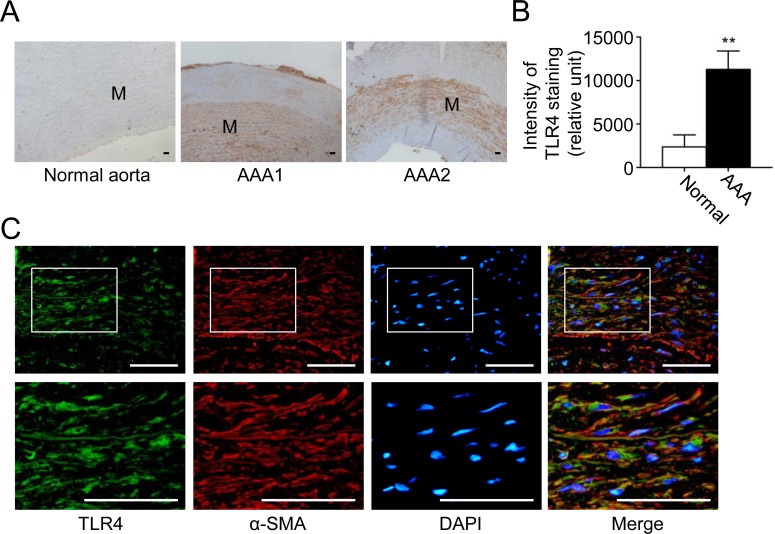
Fig 5. TLR4 is highly expressed in human AAA.
(A) Representative microscopic photos of TLR4 expression (stained red-brown) in the human normal aortas and AAAs. (B) TLR4 staining in VSMCs in human normal aortas and AAAs (n = 8 per group). (C) Representative microscopic photos of double-immunofluorescent staining for TLR4 with α-SMA in AAA. (**P<0.01 compared with normal aorta group. M indicates media. Scale bars represent 100 μm [A] and 50 μm [C].)

