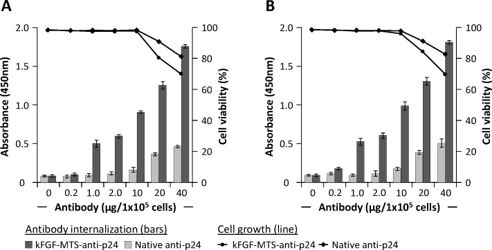
Fig 2. Concentration -dependent internalization of κFGF-MTS-anti-p24 mAb into T cells.
Cells were incubated with indicated concentrations of κFGF-MTS-anti-p24 mAbs or native anti-p24 mAb at 37°C for 18 h. Cells were treated with Trypsin and washed to remove non-internalized antibodies. Washed cells were homogenized and clarified cell lysates were subjected to antibody capture ELISA for the determination of antibody internalization. Bars show antibody internalization into cells whereas lines show cell viability. The results are representative of three independent experiments each done in duplicate. (A) Antibody capture ELISA and cell viability on Jurkat T cell lysates. (B) Antibody capture ELISA and cell viability on H9 T cells.

