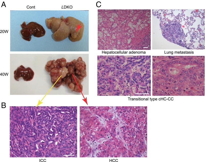
Fig. 2.
Liver tumors observed in LMob1DKO mice. (A) Macroscopic views of representative livers from 20- or 40-wk-old control and LDKO mice (n = 7). Red arrows, HCC; yellow arrow, ICC. (B) H&E-stained ICC and HCC from a 40-wk-old LDKO mouse in A. (Scale bar, 50 μm.) (C) H&E-stained tumor types in LDKO livers: hepatocellular adenoma (15-wk-old mouse); transitional type cHC-CC (40 wk); and lung metastasis (40 wk). (Scale bars, 50 μm.)

