Fig. 3.
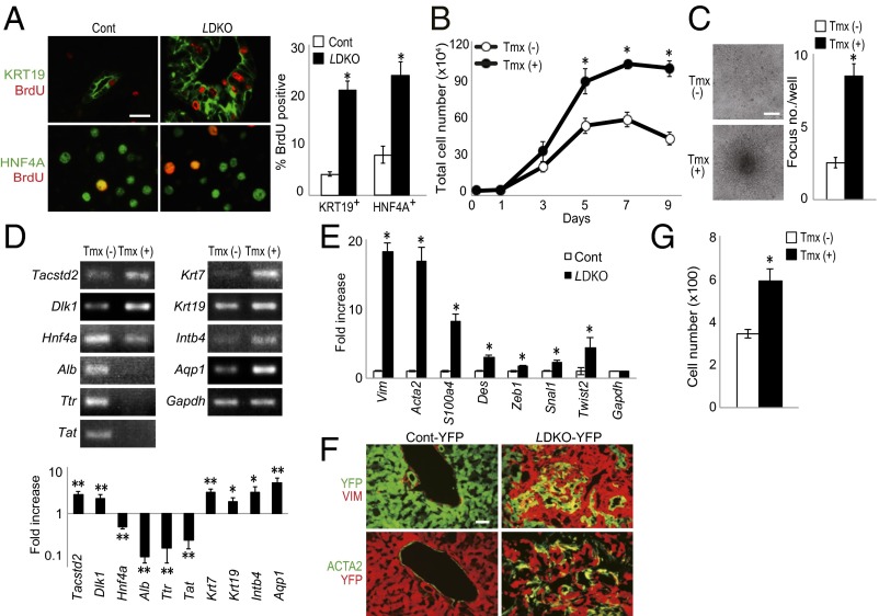
Tumorigenic anomalies in liver cells of LMob1DKO mice. (A) (Left) Immunostaining to detect BrdU incorporation (red) in P10 control and LDKO livers at 1 h post-BrdU injection. Cells were counterstained (green) with (Top) anti-KRT19 or (Bottom) anti-HNF4A Ab. (Scale bar, 20 μm.) (Right) Percentage of BrdU+ cells among the KRT19+ or HNF4A+ cells in the left panel. Data are the mean ± SEM (n = 5). *P < 0.01. (B) Total numbers of imMob1DKOLP cells cultured for the indicated number of days ± Tmx (1 μM). Data are the mean ± SEM of triplicate cultures. *P < 0.01. (C) (Left) Morphology of imMob1DKOLP cells cultured for 7 d in the absence [Tmx (−)] or presence [Tmx (+)] of Tmx (1 μM). Multiple foci are visible in the imMob1DKOLP+Tmx culture. (Scale bar, 200 μm.) (Right) Number of foci per well in the cultures in the left panel. Data are the mean ± SEM (n = 11). *P < 0.01. (D) RT-PCR (Upper) and mRNA levels (Lower) of the indicated markers in imMob1DKOLP cells cultured ± Tmx for 10 d. Tacstd2 and Dlk1, markers of liver stem/progenitor cells; Hnf4a, Alb, Ttr, and Tat, mature hepatocyte markers; Krt7, Krt19, Intb4, and Aqp1, cholangiocyte markers. *P < 0.05, **P < 0.01. (E) mRNA levels of the indicated EMT-related genes in P14 control and LDKO livers. Data are the mean ± SEM (n = 4). *P < 0.01. (F) Immunostaining to detect the indicated mesenchymal cell marker proteins in YFP+ epithelial cells in liver sections from P14 AlbCre;Mob1a+/+;Mob1b−/−;Rosa26-LSL-YFP (Cont-YFP) mice and AlbCre;Mob1aflox/flox;Mob1b−/−;Rosa26-LSL-YFP (LMob1DKO-YFP) mice (n = 4). (G) Cell migration assay of imMob1DKOLP cells ± Tmx (1μM) determined using a Transwell assay. The number of cells that had migrated into the bottom chamber after 24 h was counted in four fields under light microscopy. Data are the mean ± SEM (n = 3 chambers per group). *P < 0.01.

