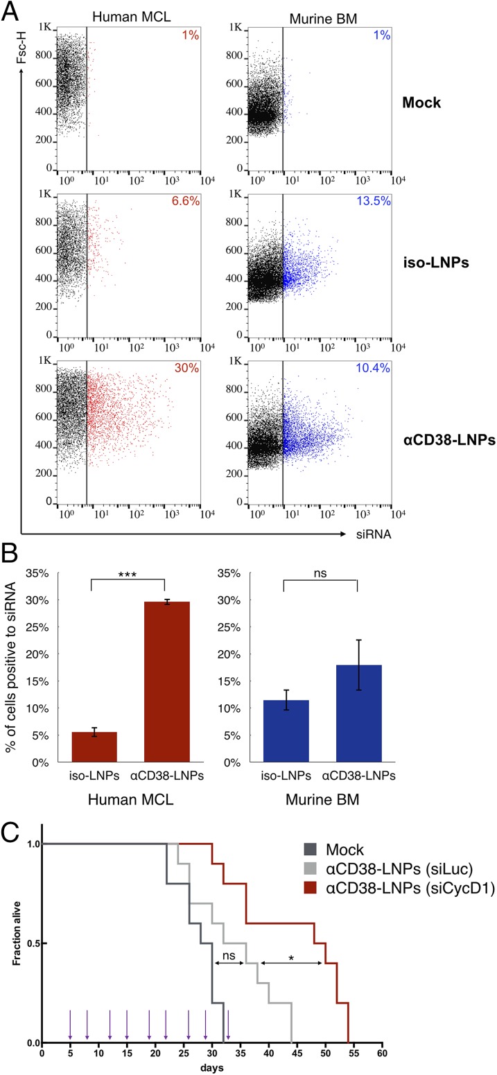
Fig. 4.
αCD38-LNPs-siCycD1 target MCL xenografts in the BM and prolong the survival of diseased mice. (A and B) Mice bearing human MCL cells were i.v.-injected with mock, isotype-, or αCD38–LNPs–siRNAs. Bone marrow cells were extracted 2 h later and analyzed for LNP binding as detected by siRNA fluorescence via flow cytometry. Human MCL (Left) and murine (Right) cells were gated separately based on GFP, hCD20, and mCD45 expression. Cells with siRNA fluorescence levels higher than in the top 1% of cells from mock-treated mice were considered positive (siRNA-positive cells are colored; cutoff is represented by the vertical bar). (A) Dot plots for one representative animal from each treatment group (isotype: n = 2; αCD38: n = 3). Number indicates percentage of siRNA-positive cells. Complete results are shown in B. Bar plots represent mean ± SEM (ns P > 0.05; ***P < 0.001; two-tailed Student’s t test). (C) Survival curves of MCL-bearing mice. Corresponding treatments (1 mg siRNA/kg body) were administrated at nine time points (arrows) via retro-orbital route. n = 10 animals per group. P values and significance were determined by log-rank Mantel–Cox test with Bonferroni correction (*P < 0.05).

