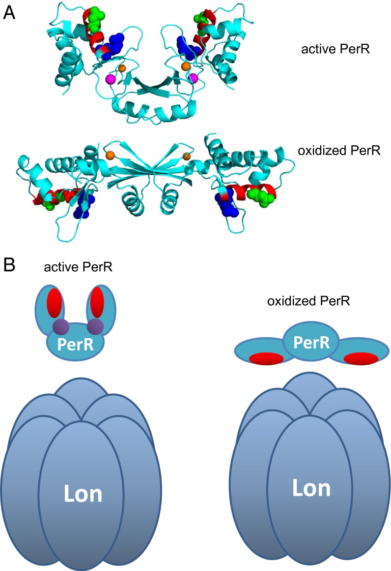
Fig. 7.
Structural position of the Lon-interacting region. (A) The helix 4 LonA-interacting region mapped on the 3D structures solved in the active (Mn-bound form, PDB ID code 3F8N; Upper) (5) and oxidized PerR (PDB ID: 2RGV; Lower) conformations (7). The α-helix (helix 4) that is the main LonA-interacting region is in red with the two critical positions colored green for N61 and blue for R67. The structural zinc is indicated in orange, and regulatory metal (Mn2+ in this case) is indicated in purple. The figure was rendered by using PyMOL (www.pymol.org) based on deposited coordinates. (B) Suggested model of substrate accessibility of PerR to LonA. PerR helix 4 is represented in red, and regulatory metal (iron or manganese) is shown in purple.

