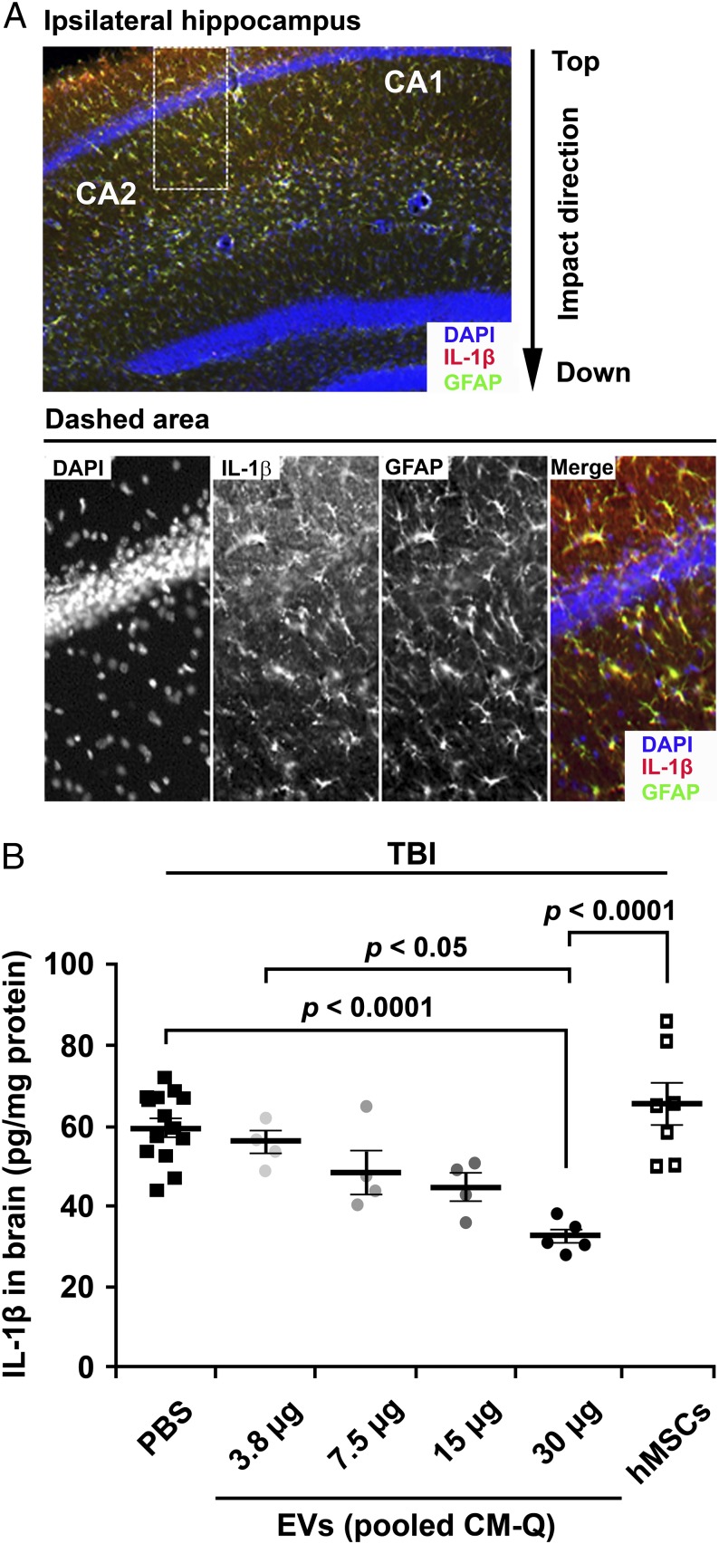
Fig. 4.
Dose–response data for suppression of neuroinflammation by EVs after TBI. (A) Immunochemistry of brain sections demonstrated that TBI increased IL-1β in GFAP+ astrocytes. Sections from brain recovered 12 h after TBI and sections from region indicated were stained for DAPI, IL-1β, and GFAP. (B) Dose-dependent decrease in IL-1β after i.v. administration of PBS or EVs. Amounts varied from 3.5 to 30 µg of protein or 1.8–15.3 × 10⁹ EVs. PBS or EVs were administered 1 h after TBI, and assays were by ELISA on homogenates of ipsilateral brain isolated 12 h after TBI. The i.v. administration of 1 million MSCs cultured in CCM had little effect, apparently because they were not fully activated in 12 h to express TSG-6 by embolization of the lung (21).

