Fig. S5.
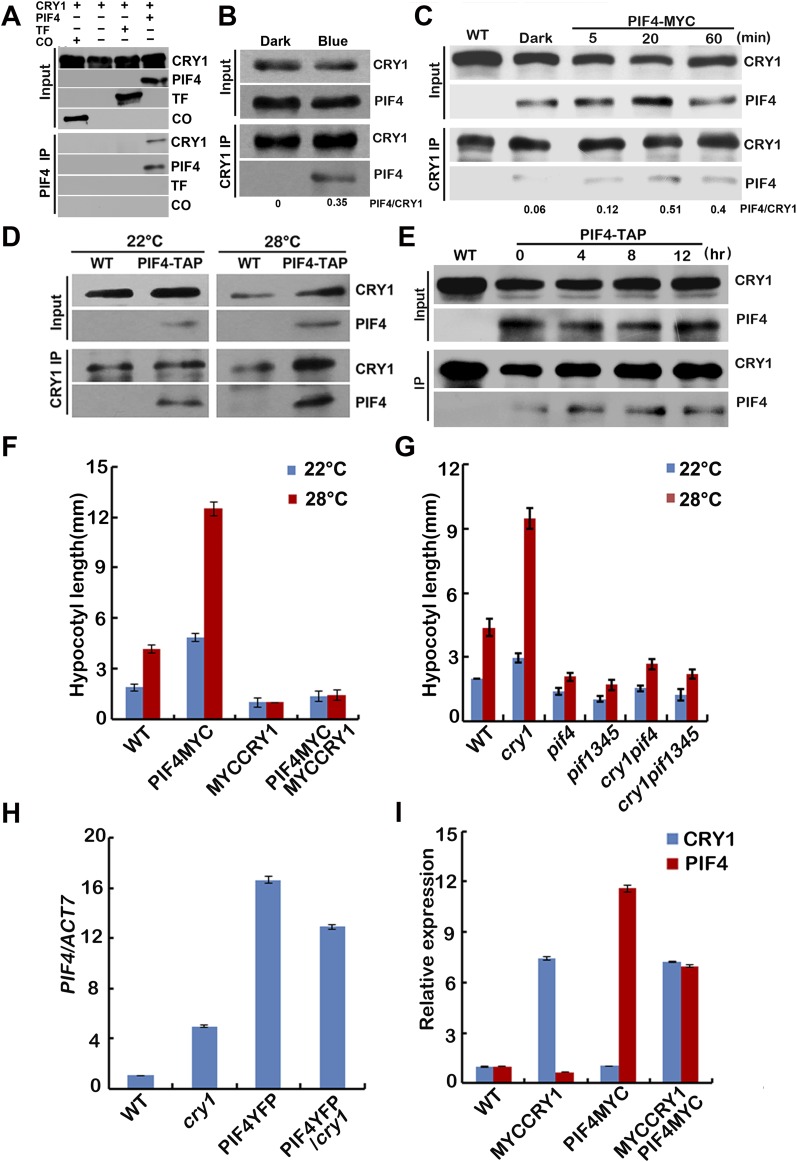
CRY1 interacts with PIF4 in a blue light-dependent manner. (A) In vitro pull-down assays showing interaction between CRY1 and PIF4. His-TF–tagged PIF4 or His-TF tag or His-TF–tagged CO (CONSTANS) were mixed with CRY1 purified from insect cells, and PIF4 antibody was used for the in vitro pull down. The products were analyzed by immunoblot probed with the anti-CRY1 (CRY1) or the anti-PIF4 antibody (PIF4) or the anti-His antibody (TF and CO). (B and C) Co-IP assays of samples prepared from 6-d-old 35S::PIF4-TAP or 35S::PIF4-MYC seedlings grown in long-day condition (16 light/8 dark), moved to dark for 1 d, then either exposed to blue light (40 μmol⋅m–2⋅sec–1) for 15 min (B) or for 5, 20, or 60 min time course (C) or kept in dark. (D and E) Co-IP assays of samples prepared from 6-d-old 35S::PIF4-TAP seedlings grown in 22 °C long-day condition, moved to 28 °C for 4 (D) or for 4, 8, or 12 h time course (E) or kept in 22 °C for 4 h. Total proteins (Input) or IP product of anti-CRY1 antibody (CRY1-IP) were probed, in immunoblots, by the anti-CRY1 antibody (CRY1), stripped and reprobed by the anti-MYC (PIF4-TAP or PIF4-MYC) antibody. (F and G) Phenotypic analysis. Seedlings of the indicated genotypes were grown in the 22 °C or 28 °C continuous white light (40 μmol⋅m–2⋅sec–1) for 4 d. The hypocotyl lengths of the indicated genotypes were measured and are shown in F and G. SDs (n > 15) are indicated. (H and I) qPCR results showing the expression of PIF4 (H) or PIF4 and CRY1 (I) in different genotypes indicated. Four-day-old seedlings grown at 22 °C in LD condition were used. Error bars represent SD of three technical repeats. Each experiment was performed at least three times with similar results.

