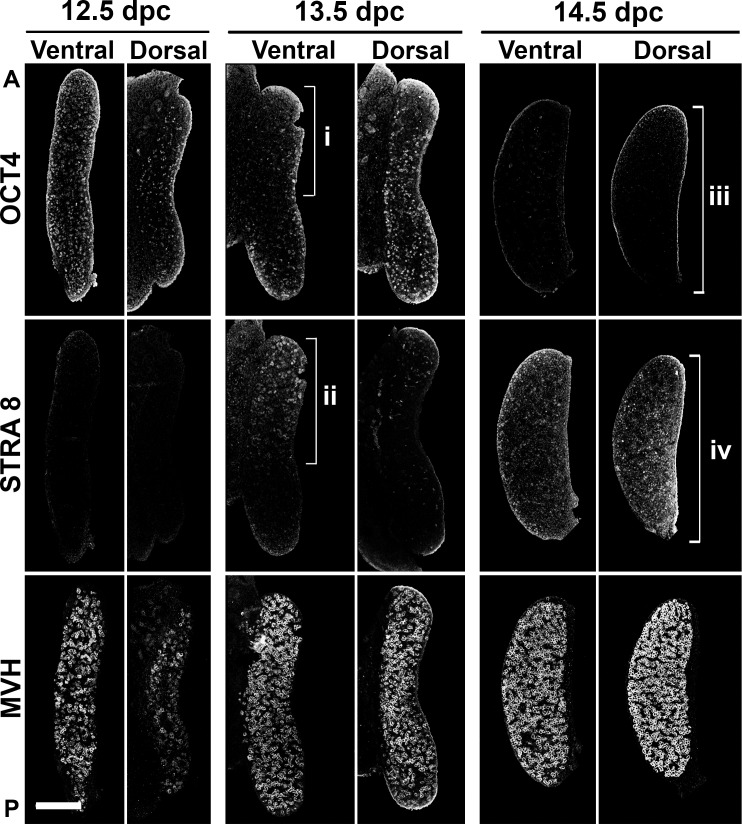
FIG. 2.
Timing of meiosis entry differed between ventral and dorsal regions of the mouse ovary. Meiosis onset was visualized using immunofluorescence assay in serial sections with OCT4, STRA8, and MVH antibodies, allowing detection of germ cells before, during, and independent of the meiotic entry, respectively. Early onset of meiosis in the anterior-ventral region at 13.5 dpc as visualized by the depletion of OCT4 (i) and expression of STRA8 (ii) is shown. At 14.5 dpc, the dorsal region of the ovary also showed evidence of meiosis (iii and iv). Note the uniform distribution of germ cells in both regions of the gonad, visualized with MVH immunofluorescence assay. 12.5 dpc, n = 11; 13.5 dpc, n = 15; 14.5 dpc, n = 9. A, anterior; P, posterior. Bar = 200 μm.

