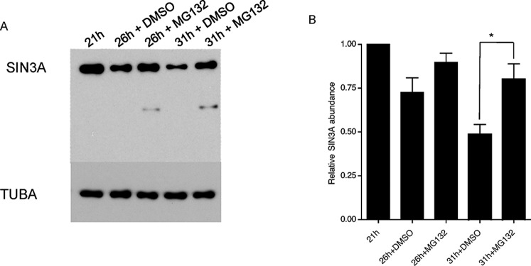
FIG. 4.
SIN3A degradation is proteasome-dependent. A) Mid-1-cell embryos were isolated and cultured in vitro in the presence of the proteasome inhibitor MG132 or DMSO, the vehicle. SIN3A abundance was measured by immunoblot analysis at the indicated times. The TUBA signal was used to normalize total protein loading. The experiment was performed three times, and a representative example is shown. B) Quantification of the data shown in A. The data are expressed as the mean ± SEM, and the SIN3A signal is relative to the mid-1-cell embryo. The times refer to the number of hours post-hCG injection. *P < 0.05.

