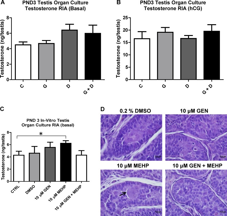
FIG. 3.
Effects of in utero and in vitro exposure to genistein (GEN) and DEHP on PND3 testes T production. Ex vivo testis organ culture was performed over 3 days in basal (A) or hCG-containing (B) medium using PND3 testes from in utero treated offspring. C) Control (untreated) PND3 testes were also treated in vitro over 3 days with either plain medium (control), vehicle (0.2% DMSO), 10 μM GEN, 10 μM MEHP, or combined 10 μM GEN + MEHP. Testosterone levels in supernatant medium for both ex vivo and in vitro organ cultures were determined by radioimmunoassay (RIA) and expressed in ng/testes. Graphs represent the sum of T produced over 3 days (supernatant collected once daily). Asterisk indicates a significant difference relative to control (P ≤ 0.05, n = 4). To assess histological alterations, testes from ex vivo (data not shown) and in vitro organ cultures (D) were collected for processing and hematoxylin and eosin staining (photos taken at 100× magnification). Arrow indicates multinucleated germ cell. Representative pictures are presented; C, control; G, GEN; D, DEHP; G + D, GEN + DEHP.

