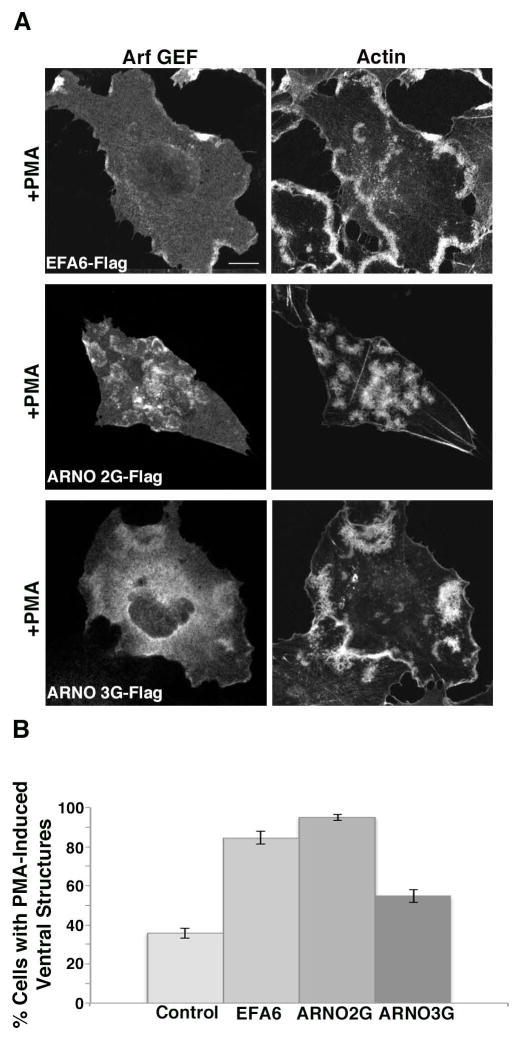
Figure 3. Arf GEFs enhance ventral actin structure formation in Beas-2b cells.
(A) Beas 2B cells were transfected with plasmids encoding Flag-EFA6 (Arf6 GEF), or the Arf1 GEFS Flag-ARNO 2G (PIP3 binding) or Flag-ARNO 3G (PIP2 binding). Cells were treated with PMA, fixed and immunostained with antibody against Flag and rhodamine phalloidin. Bar, 10 μm. (B) Percentage of cells exhibiting one or more ventral actin structures was quantified. With the exception of Flag-EFA6, in total, 200 cells were counted from three independent experiments. For Flag-EFA6, in total, 123 cells were counted from three independent experiments. Error bars represent standard error for proportional data, P<0.0001 as determined by Fisher’s Exact test (two-sided) indicating that GEF expression resulted in increase in cells with ventral actin structures.

