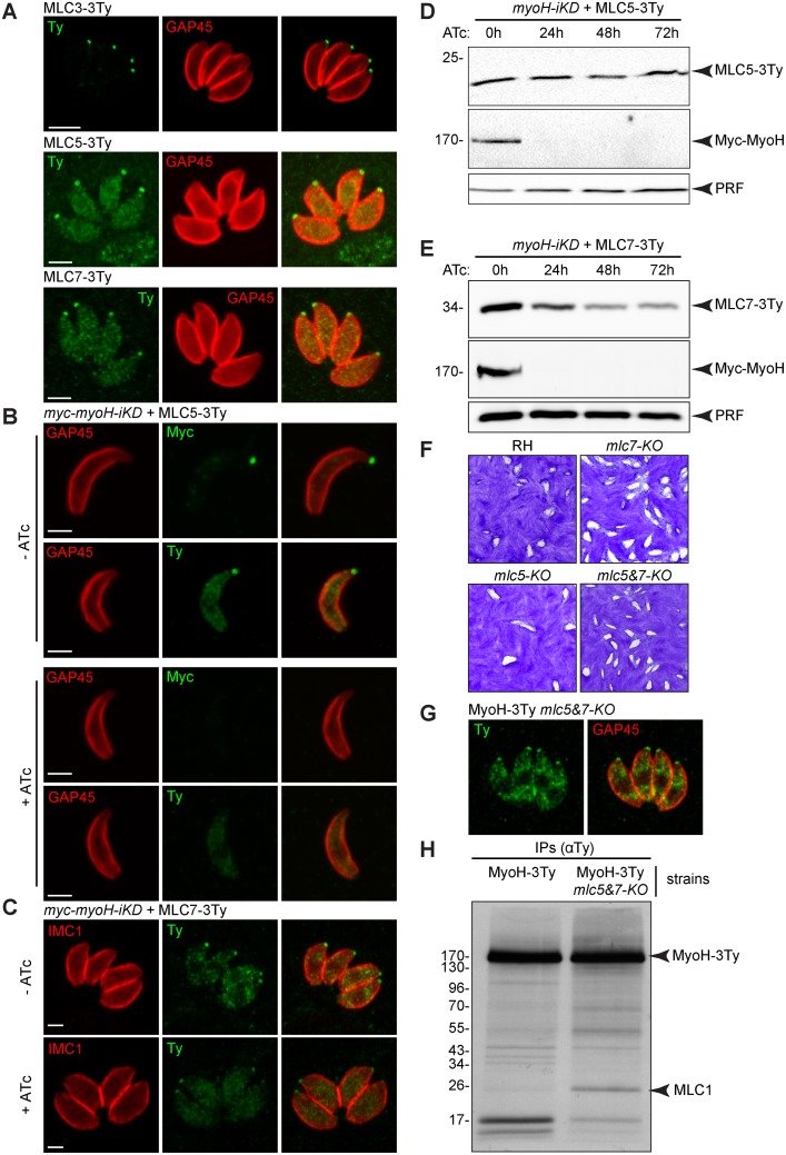
Fig 5. T. gondii myosin H is associated with three myosin light chains.
(A) Endogenously tagged myosin light chains (MLC) 3, 5 and 7 localized to the conoid. Scale bar 2 μm. (B) MLC5 localization at the conoid is dependent upon the presence of TgMyoH. Scale bar 2 μm. (C) MLC7 localization at the conoid is independent on the presence of TgMyoH. Scale bar 2 μm. (D) No decrease of the MLC5 level was observed by western blot upon depletion of TgMyoH. Profilin (PRF) serves as loading control. (E) A decrease of the MLC7 level was observed by western blot upon depletion of TgMyoH. Profilin (PRF) serves as loading control. (F) Plaque assays performed with RH (parental strain), mlc5-KO, mlc7-KO and mlc5&7-KO lines and fixed after 7 days showed no defect in the lytic cycle. (G) TgMyoH localization at the conoid is independent on the presence of MLC5 and MLC7. (H) Metabolic labeling followed by Co-IP experiments performed with anti-Ty antibodies on MyoH-3Ty and MyoH-3Ty-mlc5&7-KO strains. A band corresponding to the size of MLC1 was detected.

