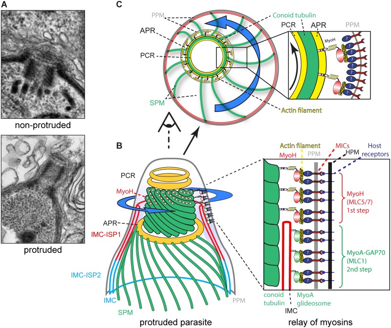
Fig 7. Model.
(A) Electron micrographs depicting the conoid in non-protruded and protruded parasites. (B) Schematic representation of the MyoH and MyoA relay at the conoid-IMC interphase. MyoH (red hoops) localized at the upper part of the conoid, close to the pre-conoidal rings (PCR). The translocation of the adhesin complexes by MyoH (blue arrow), probably via actin, are further relayed at the level of the IMC by MyoA-GAP70 in the cap region and then by MyoA-GAP45 along the rest of the parasites (see Fig 6C). APR, apical polar ring; SPM, subpellicular microtubules; PPM, parasite plasma membrane. (C) Upper view of the conoid with MyoH initiating the translocation of the adhesin complexes in a possible corkscrew-like trajectory which is likely relayed at the level of the subpellicular microtubules indirectly connected to the MyoA-glideosome.

