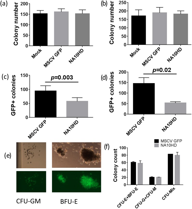
Fig 4. CFU-C assay of human CD34+ cells from four donors transduced with control GFP vector or NA10HD.
(a) and (b) showing similar total colony numbers from CD34+ cells in culture 1 day and 10–14 days post transduction, (n = 7), (c) and (d) show the GFP positive colony numbers are lower in the NA10HD group compared to MSCVGFP for cells in culture 1 day and 10–14 days post transduction, (p = 0.003 and 0.02 respectively, paired t-test, n = 7), (e) Colonies (CFU-GM and BFU-E) obtained after NA10HD transduction, (f) Similar number of erythroid colonies between MSCVGFP and NA10HD groups suggesting that erythroid differentiation is not blocked by NA10HD expression (n = 2).

