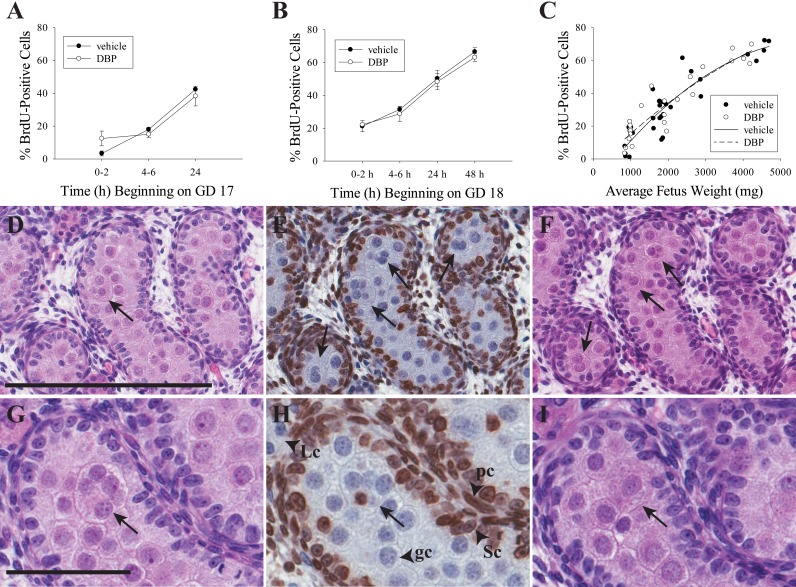
FIG. 4.
MNGs do not form by a proliferative mechanism. DBP treatment did not increase or decrease the frequency of BrdU-positive cells in the rat fetal testis when treatment began on GD 17 (A) or GD 18 (B). These values are shown in separate panels because each panel has a different start time for BrdU exposure on GD 17 and GD 18, respectively. Values in A and B are mean ± SEM. Data analyzed by two-way ANOVA followed by the Holm-Sidak test. While there were no differences by treatment within time point, the time factor was significant (P < 0.001) for GD 17 and GD 18 rats. When all samples were analyzed by fetus weight (C), the frequency of seminiferous cords with BrdU-positive cells in both vehicle- and DBP-treated fetal rat testis increased according to a quadratic trend (DBP: n = 25, R2 = 0.8574, P < 0.0001; vehicle: n = 30, R2 = 0.8307, P < 0.0001). D–F) Three serial 5-μm sections from a rat treated with DBP for 48 h beginning on GD 18, stained with H&E (D, F) or by IHC for BrdU with hematoxylin counterstain (E). Four MNGs are clearly visible in the BrdU-labeled section (all are BrdU negative). One is visible on the adjacent section D, while three are also visible on adjacent section F. A similar series of three 5-μm sections is shown in G–I, with an MNG visible in all three sections that has a single BrdU-labeled nucleus in H. In all panels, arrows indicate MNGs. Labeled arrowheads in H demonstrate normal pattern of BrdU staining in peritubular cells (pc) and Sertoli cells (Sc) but not in germ cells (gc) or Leydig cells (Lc). Bar in D = 200 μm for D–F, and bar in G = 60 μm for G–I.

