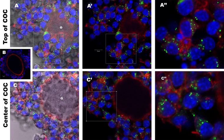
FIG. 2.
Cumulus cells uptake follicular fluid EVs during COC culture. Mouse COCs were cultured 16 h with PKH67-labeled bovine follicular EVs. Individual COCs were imaged on a Zeiss Pascal confocal microscope (×40) in sections 1 μm thick and examined for internalization of follicular EVs. In the expanded COC, internalized follicular EVs are evident as green spots within cumulus cells in both the outer layers of cumulus cells (A) and inner layers adjacent (C) to the oocyte (o). As negative control to insure that the COC labeling was a result of EV uptake and not residual free-floating dye in the media, the final supernatant (wash) from the follicular EV PKH67 labeling process was added to COC cultures, and these cumulus cells were all negative for PKH67 (B). Distance between the sections in A and C is 30 μm within the same COC. A′ and C′ are the same as A and C but with DIC turned off. A″ and C″ are enlargements from the boxes identified in A′ and C′. Bar = 10 μm.

