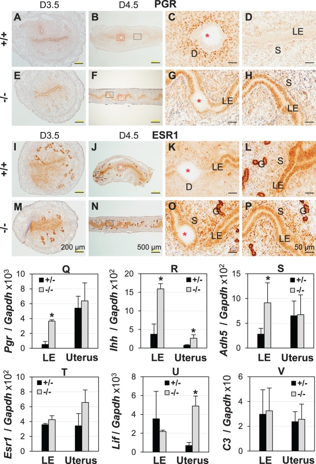
FIG. 3.
Uterine expression of PGR and estrogen receptor α (ESR1) and their target genes. D3.5/D4.5, Gestation Day 3.5/4.5; +/+, Lpar3+/+; +/-, Lpar3+/−; −/−, Lpar3−/− mice. A–H) Immunohistochemistry (IHC) of PGR. A) Cross-section of a uterus, +/+, D3.5. B) Longitudinal section of a uterus, +/+, D4.5. C) Enlarged view of the red rectangular area in B. D) Enlarged view of the black rectangular area in B. E) Cross-section of a uterus, −/−, D3.5. F) Longitudinal section of a uterus, −/−, D4.5. G) Enlarged view of the red rectangular area in F. H) Enlarged view of the black rectangular area in F. I–P) IHC of ESR1. I) Cross-section of a uterus, +/+, D3.5. J) Longitudinal section of a uterus, +/+, D4.5. K) Enlarged view of the red rectangular area in J. L) Enlarged view of the black rectangular area in J. M) Cross-section of a uterus, −/−, D3.5. N) Longitudinal section of a uterus, −/−, D4.5. O) Enlarged view of the red rectangular area in N. P) Enlarged view of the black rectangular area in N. Bars = 200 μm (A, E, I, M), 500 μm (B, F, J, N), 50 μm (C, D, G, H, K, L, O, P). Q–V) Quantitative PCR of mRNA expression in D4.5 LE and uterus. Q) Pgr. R) Indian hedgehog (Ihh). S) Alcohol dehydrogenase 5 (Adh5). T) Esr1. U) Leukemia inhibitory factor (Lif). V) Complement component 3 (C3). *P < 0.05; n = 3; error bars, standard deviation; glyceraldehyde 3-phosphate dehydrogenase (Gapdh), loading control.

