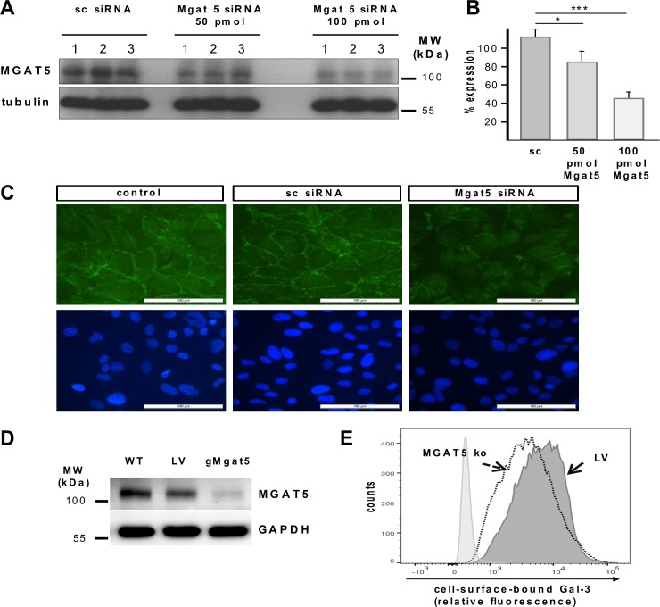
Fig 4. Knockdown of Mgat5 expression in cultured human RPE cells attenuates binding of Gal-3 to RPE cells.
(A) Western blot analysis of Mgat5 expression in RPE cells transfected with siRNA containing the same nucleotides as Mgat5 siRNA in random order (sc siRNA), and the same cell line transfected with 50 pmol and 100 pmol of double stranded siRNA complementary to Mgat5 (Mgat5 siRNA), respectively. Lysates containing approximately equal amounts of protein were separated by SDS-PAGE and blotted for immunochemical detection of Mgat5 content. Experiments were repeated at least three times. MW; molecular weight. (B) Quantification of Mgat5 gene silencing. Values are normalized to expression of tubulin. (C) Fluorescence micrographs of Gal-3 binding to the RPE cell surface. Ninety-six hours after transfection cells were treated with 60 μg/mL biotinylated Gal-3. Cells were then fixed and stained with a fluorescent streptavidin conjugate. Nuclei were counterstained with DAPI. Localization of Gal-3 binding was visualized by fluorescence microscopy at a 40 fold magnification. Scale bars represent 100 μm. Untreated cells exposed to streptavidin conjugate alone served as negative controls and exhibited no fluorescence signal (data not shown). (D-E) The target sequence derived from the genomic sequence of Mgat5 was inserted into a CRISPR-Cas9 nuclease expressing lentiviral vector and ARPE19 cells were transfected using Lipfectamine 2000 Plus reagent. (D) Western blot analysis of Mgat5 expression in ARPE19 cells transduced with guide RNA leading to specific knockdown of the Mgat5 (gMgat5), or cells tranduced with a CRISPR-Cas9 lentiviral vector encoding for an none-coding filler RNA (LV), or wild-type ARPE-19 cells. Lysates containing approximately equal amounts of protein were separated by SDS-PAGE and blotted for immunochemical detection of Mgat5 content. (E) Flow cytometric analysis of Gal-3 binding in Mgat5-knockout cells. CRISPR-Cas9-mediated Mgat5 knockdown of cultured RPE cells reduces cells surface binding of Gal-3, when compared to cells transfected with a non-coding control vector alone (LV). Histograms represent the number of counted cells versus relative fluorescence intensity. Transduction experiments have been repeated three times.

