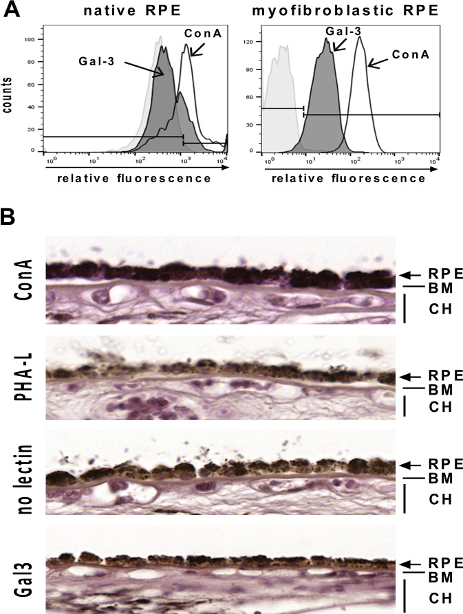
Fig 6. Native RPE cells exhibit little binding of Gal-3.
(A) Flow cytometric analysis of Gal-3 binding to native and myofibroblastic RPE cells. Binding of Gal-3 (gray) to native RPE cells was only slightly above background, whereas Gal-3 binding to myofibroblastic RPE cells was evident. ConA binding was markedly above background in both cell populations. Histograms represent the number of cells versus relative fluorescence intensity. Experiments have been repeated two times. (B) Lectin histochemistry of human RPE cells in situ. Human cadaver eyes were incubated with biotinylated lectins as indicated on the left and binding was visualized by incubation with streptavidin-coupled peroxidase and Vector VIP substrate™ (purple color). In control sections with the VIP substrate alone the RPE can be easily discerned by the characteristic brownish pigment (third panel from the top). RPE cells and extracellular matrix reacted strongly with ConA (top panel), whereas PHA-L (second panel from the top) did not recognize native RPE cells and exhibited a staining pattern comparable to that of substrate alone. Biotinylated Gal-3 (purple color, fourth panel) did not bind to native RPE in situ and staining patterns resembled closely the situation in untreated negative control eyes. RPE, retinal pigment epithelium; BM, Bruch´s membrane; CH, choriocapillaris.

