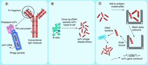
Figure 8. . Creation and selection of antibody phage display libraries.
(A) Cartoon depicting a prototypical IgG immunoglobulin and corresponding antibody-displaying filamentous phage particle. IgG comprises a heavy chain variable region (VH) and 3 constant region domains (CH1, CH2 and CH3). The IgG light chain comprises a variable region (VL) and one constant region domain (CL). An Fv fragment is defined as the combination of heavy and light chain variable regions only (VH + VL). For simplicity in recombinant expression of Fv fragments, the VH and VL domains can be expressed as a single polypeptide separated by a short flexible glycine/serine-rich linker referred to as a single-chain Fv (scFv). Arrows on the IgG variable region represent the positions of forward and reverse polymerase chain reaction primers that are used to amplify the heavy and light chain gene segments. A second PCR step links the VH and VL into one genetic construct that can ligated into a phagemid expression vector that appends the gene for the phage pIII coat protein to the carboxy terminus of the scFv as depicted on the phage particle. (B) Cartoon depicting the construction of an antibody phage display library from a pool of B lymphocytes. Phage particles each express an scFv on their surface that is encoded by the DNA contained within the particle. Arrows indicate a potential correspondence between a particular B cell and a phage particle displaying that B cell's immunoglobulin. (C) Schematic representation of phage panning procedure in which an aliquot of phage library is incubated with immobilized antigen and adsorbed phage are eluted and amplified in bacterial culture (see text for details).

