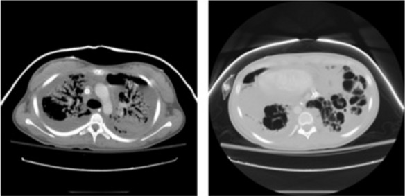
Figure 1.

a) Axial image from a chest CT showing severe bilateral upper lobe consolidation with numerous air bronchograms, a loculated left hydropneumothorax and a loculated right pleural effusion. An ECMO cannula in the SVC is noted. b) A more caudal axial image demonstrates widespread cystic destruction of the lower lobe parenchyma representing necrosis. CT = computed tomography; ECMO = extracorporeal membrane oxygenation; SVC = superior vena cava.
