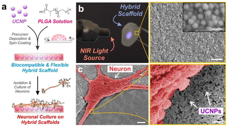
Figure 3. Blue-emitting UCNPs were embedded within thin polymer films of PLGA and cultured with hippocampal neurons.
(a) Schematic depicting preparation of poly(lactic-co-glycolic acid) (PLGA)-embedded UCNP hybrid scaffolds for hippocampal neuronal cultures. (b) Photographic image of the flexible hybrid scaffold film. The blue luminescence under 980-nm laser excitation illustrates the encapsulation of UCNPs throughout the PLGA film. Scanning electron microscopy (SEM) image shows the distribution of the UCNPs within the PLGA film. Scale bar: 500 nm (bottom), 100 nm (inset). (c) Scanning electron microscopy (SEM) image of a hippocampal neuron (pseudo-colored red for contrast) cultured on the polymer-UCNP hybrid scaffolds at 14 DIV. Scale bar: 5 μm (left), 200 nm (right).

