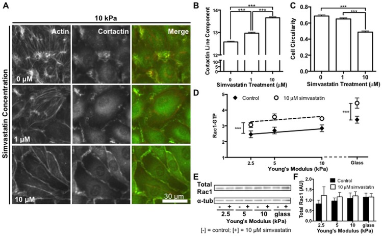
Fig 4. Simvastatin alters actin organization, cell morphology, and Rac1 activity in endothelial monolayers.
Cytoskeletal organization and Rac1 activity in endothelial monolayers is altered by simvastatin treatment. (A) Representative images of endothelial monolayers showing prominent actin stress fibers in control cells and a barrier enhancing cortical actin ring that forms with increasing simvastatin concentration. Cortactin changes from puncta to organized linear segments around the cell periphery and localizes with actin with increasing statin treatment. (B) Cortactin organization, measured by quantifying linear segments at cell-cell junctions, increases with simvastatin (n = 3, 30 fields of view per condition). (C) Endothelial cells adopt an elongated morphology as actin stress fibers diminish with increasing simvastatin treatment. Cell circularity, where a perfectly circular cell has a value of 1, decreases with increasing simvastatin concentration (n = 3, 50–54 cells per condition). (D) Rac1-GTP activity normalized to total protein of lysate increases across all stiffness levels with simvastatin treatment (n = 5, performed in duplicate or triplicate). (E) Representative Western blot probing for total cellular Rac1 expression and alpha tubulin (α-tub) loading control. (F) Quantification of total Rac1 normalized to alpha tubulin loading control demonstrating no significant change in expression with stiffness or statin treatment (n = 5). Data are presented as means ± standard error of the mean, ***p<0.001.

