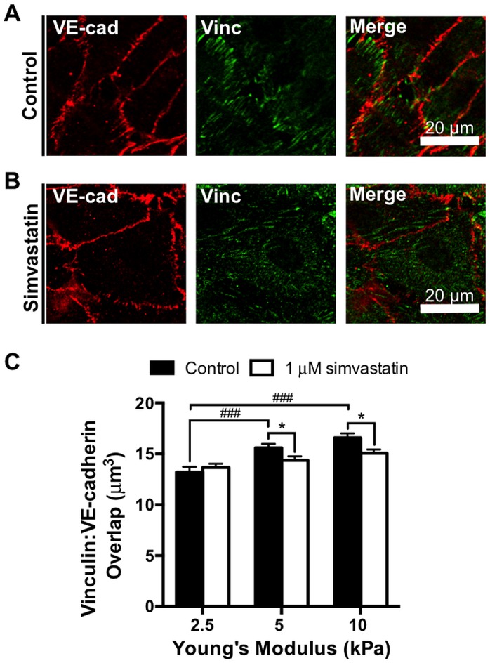
Fig 7. Simvastatin reduces vinculin localization at cell-cell adhesions in endothelial monolayers.

Endothelial monolayers treated with control or 1 μM simvastatin are stained for vinculin and VE-cadherin, and the vinculin volume overlapping with VE-cadherin is quantified. Representative images of endothelial cell-cell junctions within a confluent monolayer fluorescently stained for vinculin and VE-cadherin on a 10 kPa polyacrylamide gel after 24 hour (A) control or (B) 1 μM simvastatin treatment demonstrating vinculin positive and vinculin negative junctions, respectively. (C) Vinculin localization per monolayer at cell-cell adhesions, a readout of intercellular junction tension, is quantified and increases with matrix stiffness but is significantly decreased with the statin treatment at higher matrix stiffnesses (n = 3, 70–90 fields of view per condition). Data are presented as means ± standard error of the mean, ###p<0.001 compared to matrix stiffness, *p<0.05 compared to the untreated control.
